Chemical Engineering Tutorials: 2024

Thursday, 5 December 2024

Entropy and the Second Law

We know that for a variety of processes, the entropy of the universe increases for an irreversible process and remains constant for a reversible process:

 (1)      

If we assume the universe to be an isolated system, we can rephrase the Second Law as

“Any spontaneous process occurring in an isolated system will always result in an increase in that system’s entropy.”

Thus:

  • For an isolated system, if an entropy increase is not possible, then a spontaneous change cannot occur in that system. This is possible if the entropy of an isolated system is at its maximum value. The condition of maximum entropy i.e., no possible spontaneous change is called the state of thermodynamic equilibrium.
  • The statement: “the entropy of a system can never decrease” only applies to an isolated system. Generally, the entropy of an arbitrary system can be decreased by a suitable heat interaction.
  • In order to determine the priority of a process occurring in the universe, we have to apply the principle of entropy increase. That is, any spontaneous process following a previous process must have a higher entropy than the preceding process. Thus, entropy is referred to as time’s arrow.

It should be noted that there is no such thing as the conservation of entropy. Real processes always generate entropy and, as a result, entropy of the universe always increases.


a) Entropy and the Loss of Work

Let us consider a rigid, well-insulated tank divided into two equal volumes A and B by a membrane. Side A is filled with 1 mol of an ideal gas at 300K and 1bar and the side B is evacuated. If the membrane ruptures so that the gas fills the entire volume, what would be the entropy change?

System: The contents of the tank.

Using the 1st law of thermodynamics:

Since the internal energy of an ideal gas is dependent only on temperature, the final temperature is also 300K. We can use the following equation to help calculate the entropy change of the system:

Thus, the entropy change of the system can be solved by simplifying the above equation. Also, since there is no heat interaction between the system and its surroundings, entropy change of the surroundings is zero. Thus:

To compare this irreversible process with a reversible one between the same initial and final states, we can consider 1 mol of an ideal gas at 300K and 1 bar in a piston-cylinder assembly. If we assume that the gas expands until the volume doubles against a frictionless piston while heat is reversibly transferred to the gas in order to keep the gas at a constant 300K. Thus the entropy change of the system is: 

The amount of heat transferred to the gas can be calculated using equation 7:


When the gas expands irreversibly in a rigid and well-insulated tank, we see that the entropy of the system increases and no work is done by the system. Conversely, reversible expansion of the gas from the same initial state to the same final state increases the entropy of the system by the same amount but, in this case, work is done by the system.

In the reversible process, the increase in the entropy of the system is compensated by the decrease in the entropy of the surroundings so that ΔSuniverse = 0. In other words, entropy is transferred from the surroundings into the system. Thus, entropy generation corresponds to the decrease in the ability of energy to do work.

If energy is a measure of system’s ability to do work, entropy is a measure of how much this ability has been devalued. Entropy generation is directly related to the lost work.


b) Entropy and Probability

One mole of a gas contains N molecules, where N is the Avogadro’s number (N = 6.02 × 10²³).

Let us consider a rigid, well-insulated tank divided into two equal volumes by a diaphragm with a hole in it and the tank contains 1 mol of an ideal gas.

Note that the probability of finding all the molecules in the tank is unity.

Hypothetically, if we capture one of the molecules and paint it red, the probability of finding that red molecule on the left side is 1/2. If we capture another molecule and paint it white, the probability of finding both the red and white molecules on the left side is (1/2)(1/2) = (1/2)2. The probability of finding all the molecules on the left side is almost zero and equal to (1/2)N

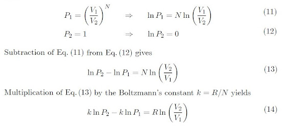
It doesn’t matter where the position of the diaphragm is, the probability of finding all the molecules on the left side is (V1/V2)N, where V1 is the volume to the left of the diaphragm and V2 is the total tank volume.

If we suppose that the gas is initially contained on the left side. By puncturing the diaphragm, the gas fills the entire tank. In this way, the gas goes from a state of low probability to a state of high probability. Let P1 and P2 represent the initial and final probabilities, respectively. Thus,

During expansion, the internal energy and the temperature of the gas remains constant.

As a result, the right-side of Equation 14, gives the change in entropy of an ideal gas during isothermal expansion from V1 to V2. Therefore, Equation 14 becomes:


Thus, the increase in entropy during a spontaneous process corresponds to the transition from a state of lower probability to a higher probability.


c) Entropy and Disorder

In a system the entropy increases with increasing disorder. Thus, the disorder in the universe is always increasing.

As the system changes to a more orderly state, its entropy decreases. Some examples are:

  • During phase transformation, i.e., Liquid to solid and gas to liquid
  • As the temperature of a substance decreases
  • As the pressure of a gas increases.

The molecules of a perfect crystal at 0 K have no internal energy, hence the molecules are perfectly ordered in the crystal lattice. 

Thus, one can assign the value of zero to the entropy for this idealized state of maximum possible order. This assignment turns out to be very useful and is sometimes referred to as the third law of thermodynamics.


Friday, 15 November 2024

Continuum Mechanics

Continuum Mechanics is the study of the behavior of materials by ignoring its particulate nature. 

A continuum is an area that can keep being divided and divided infinitely with no individual particles. It is a simplification that allows us to investigate the movement of matter on scales larger than the distances between particles.

In continuum, the smallest element of a fluid is NOT a fluid molecule, but rather a fluid particle that contains enough number of molecules to make meaningful statistical averages. Continuum assumes that fluid and flow properties like pressure, temperature, density, velocity, etc. vary continuously throughout the fluid.

This helps to study a wide range of phenomena, from air and water flow to even the evolution of galaxies.

For us to know whether or not the continuum hypothesis can be used, a dimensionless number called Knudsen number is used. The Knudsen number allows for characterizing the boundary conditions of a fluid flow. It is defined as:


If the length scale of the fluidic system is in the same range as the mean free path, i.e., Kn = 1, the fluid cannot be treated as a continuum.

The Knudsen number is very useful when assessing the boundary of fluid flows. Usually, the flow at the boundary of a flow field where the channel walls are fixed in space and the liquid directly in contact is considered to not be moving. This is referred to as the no-slip boundary condition, i.e., there is no relative movement (slip) between the wall and the fluid layer that directly contacts with the wall. 

According to the Knudsen number, flows can be divided into different regimes:


(a) For Kn < 0.01, continuum flow dominates and conventional fluid dynamics equations are applicable, indicating that gas molecules interact with neighboring molecules.

(b) For 0.01 < Kn < 0.1, the slip flow regime occurs when the gas molecules experience slipping at the solid interface.

(c) For 0.1 < Kn < 10, transition flow occurs. This refers to a flow regime in which both slip (continuum) and diffusion flows can occur.

(d) For Kn > 10, Knudsen's (free molecular) flow occurs. This refers to gas molecules that flow with minimal or no interaction with neighboring molecules.


Fluid Properties:

Characteristics of a continuous fluid which are independent of the motion of the fluid are called basic properties of the fluid. Some of the basic properties are as discussed below:

  • Density: The density ρ of a fluid is its mass per unit volume (kg/m3).
  • Specific Weight: This is the weight of a fluid per unit volume (N/m3).
  • Specific Volume: is the volume occupied by unit mass of fluid (m3/kg).
  • Specific Gravity: For liquids, it is the ratio of density of a liquid at actual conditions to the density of pure water at 101 kN/m2, and at 4°C.
    • For gases, the specific gravity is the ratio of its density to that of either hydrogen or air at some specified temperature or pressure.


Saturday, 2 November 2024

First and Second Law of Thermodynamics Solved Questions

Note: For the following examples, Appendix B for steam values that I have referred to were obtained from: 

M.D Koretsky, Engineering and Chemical Thermodynamics, Wiley, 2004.

Example 1

2 Kgs of steam at 800 kPa and 600°C is contained in a piston-cylinder assembly. Calculate the work if:

a) The steam expands reversibly and isothermally to 300 kPa.

b) The steam expands reversibly and adiabatically to 300 kPa.

Solution

System: Contents of the piston cylinder assembly

a) The work done by the system is calculated as follows:


It is necessary to plot a P versus V graph and calculate the area under the curve. 

Since expansion occurs at T = 600°C, then the variation of V as a function of P can be determined from the steam table as follows:

The plot of P versus V is shown in the figure below. (Plotted using MathCAD)

The area under the curve can be solved using the trapezoidal rule:

Alternatively, the work done by the system can be solved using the 1st law of thermodynamics

From Appendix B


b) The 1st law of thermodynamics simplifies as:

Since the process is reversible and adiabatic (or isentropic), the entropy remains constant at a value of 8.1332 kJ/kg.K. Therefore, the properties at the final state are:

Substituting these values into equation3:


Example 2

Consider the system shown in the figure below:

The piston is made of a non-heat-conducting material and the tank is insulated. Initially, the piston is at the extreme left-hand-side of the cylinder and the cylinder contains 1.5 kg of steam at 200°C and 100 kPa. The cylinder is connected to an infinite source of air at 500 kPa and 250°C, and the valve is opened slightly until the pressure in the cylinder reaches 500 kPa.

a) Calculate the final temperature of the air under these conditions.

b) Calculate the final temperature of the air if a small cooling coil is placed in the cylinder to maintain the steam temperature constant at 200°C.

Solution

Let the subscripts A and S stand for air and steam, respectively.

Since steam occupies the whole volume of the cylinder initially, the volume of the cylinder is calculated from the initial conditions. From Appendix B (Table B.4)

Thus:

a) The steam in the cylinder undergoes a reversible and adiabatic compression,


b) In this case, the steam in the cylinder undergoes a reversible and isothermal compression. Thus, the properties of the steam at the final state are:


Considering the air in the cylinder as a system, the unsteady-state mass and energy balances again lead to:


Tuesday, 15 October 2024

Reactor Sizing Examples

 Reactor sizing was discussed in a previous blog entry (Click here to view).

Example 1

Consider a liquid phase reaction occurring in a PFR with the following data and Equation

X

0

0.4

0.8

-rA (mol/dm3s)

0.01

0.008

0.002

1/(-rA) (dm3s/mol)

100

125

500


If the molar feed to the PFR is 2 mol/s, what is the volume of the PFR needed to achieve 80% conversion.

Answer

For a PFR, 

Using the data provided in the table, we can plot a Levenspiel Plot:


Using Simpson's three point rule:


Thus, to achieve an 80% conversion, a PFR of  293.3 dmis needed

Example 2

A second order irreversible reaction is carried out in the gas phase inside a PFR. 

A reactant of molecular weight 40 and 50% by weight, and the rest with an inert of molecular weight of 20 are fed to the reactor. The reaction is carried out at constant temperature of 70°C and constant pressure of 5.25 atm. The rate constant is 400m3/(kmol.ks).

Calculate the volume of the PFR needed to achieve a 40% conversion of A to produce 30kmol/hr of product R.

Answer

For a second order reaction:

When substituting into the PFR equation and integrating the equation we get:

            (1) 

To calculate εA, let us consider a 1.0g sample of reagent mixture which has 0.5/40 = 0.0125 mol of A and 0.5/20 = 0.025 mol of inert. The following volume balance is then obtained. 


The expansion parameter, εA, = (0.05 - 0.0373)/0.0373 = 0.0340

The initial concentration of A, CA0, is given by:

Substituting into equation 1,


Given the product flow rate (FR) is 30 kmol/hr, we can obtain the volumetric flow rate: 

The reactor volume can now be found using the following:


Tuesday, 24 September 2024

Scanning Electron Microscopy

Scanning Electron Microscopy (SEM) is an extremely useful technique used to obtain high-resolution images and detailed information of a samples’ surface. The typical resolution of SEM instruments ranges from < 1 nanometer to several nanometers. SEM uses a focused beam of electrons to examine the surface of a sample and analyzes the different signals emitted in a detector.

The following is an illustration of how an SEM instrument works:

Image Source: https://www.technologynetworks.com/analysis/articles/sem-vs-tem-331262

1) Electron generation. 

A high-voltage electron gun (typically 1 keV – 30 keV) generates and accelerates a beam of electrons, typically using thermionic or field emission.

  • Thermionic emission involves heating a thin tungsten filament to a high temperature (approximately 2,500°C), causing electrons to be emitted. 
  • Field emission is an advanced technology used to capture the microstructure image of a material. It is usually performed in a high vacuum because gas molecules tend to disturb the electron beam and the emitted secondary and backscattered electrons used for imaging. Field emission produce electron beams with the highest brightness and coherence which allows for the highest resolutions to be obtained.

2) Electron beam focusing

The electron beam generated is then focused on the sample using a series of electromagnetic lenses. These lenses are similar to optical lenses, but instead of glass lenses, they use magnetic fields to bend the electron beam.

Condenser lenses are used for this and are carefully aligned to ensure that the beam is focused to a small spot on the sample surface.

3) Scanning

The focused electron beam is scanned across the sample surface in a raster pattern, similar to how a television screen is scanned. 

This is achieved by using scanning coils that deflect the beam in the x and y directions.

4) Interaction of electron beam and the sample

As the electron beam interacts with the sample, it results in a variety of signals being emitted. These signals include:

  • Secondary electrons: Low-energy electrons that are emitted from the sample as a result of the primary electron beam striking the surface.
  • Backscattered electrons: High-energy electrons that are reflected back from the sample. These are high-energy electrons used to obtain high-resolution images that show the distribution of various elements that make up a sample.
  • Characteristic X-rays: X-rays that are emitted when the primary electrons knock out inner-shell electrons from the atoms in the sample. The energy difference between two electron shells is equal to the characteristic X-ray energy. Characteristic X-rays that result from inelastic scattering allow for exact identification of elements present in the sample.
  • Auger electrons: These are emitted at discrete energies that are characteristic of the elements present on the sample surface.  The characteristic energies of the Auger electrons are such that only the electrons from the outer 0.5 to 5 nm of the sample can escape and be detected.

5) Detection of emitted signal

The emitted signals are detected using various detectors, such as:

  • Secondary electron detector: Detects secondary electrons, which are used to create an image of the sample's surface topography.
  • Backscattered electron detector: Detects elastically scattered electrons, which can provide information about the sample's composition.
  • X-ray detector: Detects characteristic X-rays, which can be used to determine the elemental composition of the sample.
  • Auger electron spectrometer: Detects Auger electrons, which can be used to analyze the surface composition of the sample.

6) Formation of image

The detected signals are processed by a computer to create an image. The brightness of each pixel in the image corresponds to the intensity of the detected signal at that point on the sample. 

SEM images can be displayed in various modes, such as bright field, dark field, and elemental mapping.

7) Analysis of image

The SEM images can be analyzed to deduce the quantitative information about the sample, like size, shape, and distribution of features. 

This can be done using image analysis software or by manually measuring features on the image.


Application of SEM

a) Materials Science

  • Microstructure analysis: SEM is widely used to study the microstructure of materials like metals, ceramics and polymers. By examining the arrangement of grains, defects, and inclusions, researchers can gain insights into the material's properties and behavior. 
  • Failure analysis: When a material or component fails, SEM can be used to investigate the root cause of the failure. By examining the fracture surface, researchers can identify the type of fracture (e.g., brittle, ductile, fatigue) and identify potential defects or flaws that may have contributed to the failure.

b) Biology and Medicine

  • Cell imaging: SEM can provide high-resolution images of cells and their organelles, allowing researchers to study their structure, function, and interactions. For example, SEM can be used to examine the surface morphology of bacteria, the structure of viruses, and the ultrastructure of tissues.
  • Tissue analysis: SEM can be used to analyze the structure and composition of tissues, such as skin, bone, and muscle. This can be helpful for diagnosing diseases, studying the effects of treatments, and understanding biological processes.

c) Nanotechnology

  • Characterization of nanomaterials: SEM is a valuable tool for characterizing nanomaterials, such as nanoparticles, nanotubes, and nanowires. It can be used to measure the size, shape, and distribution of nanomaterials, as well as to study their surface morphology and internal structure.

d) Other Fields

  • Forensic science: SEM can be used for various forensic applications, such as analyzing fingerprints, examining crime scene evidence, and identifying fibers or hairs.
  • Semiconductor manufacturing: SEM is essential for quality control in the semiconductor industry, as it can be used to inspect the surface features of wafers and devices.
  • Environmental science: SEM can be used to study the morphology and distribution of pollutants in the environment, such as particulate matter and microplastics.


Advantages of SEM

  • High Resolution: SEM can provide images with extremely high resolution, allowing for detailed examination of even the smallest structures.
  • Depth of Field: SEM has a large depth of field thus it can focus on a wide range of depths simultaneously, making it ideal for imaging rough or uneven surfaces.
  • Three-Dimensional Imaging: SEM can create 3D images of samples, providing valuable insights into their structure and morphology.
  • Versatile Applications: SEM can be used to study a wide range of materials and samples, from metals and ceramics to biological tissues and nanomaterials.
  • Quantitative Analysis: SEM can be used to obtain quantitative information about samples, such as the size, shape, and distribution of features.

Disadvantages of SEM

  • Sample Preparation: SEM requires careful sample preparation, often involving coating the sample with a conductive material to prevent charging. This can be time-consuming, expensive and may introduce artifacts.
  • Vacuum Environment: SEM operates in a vacuum environment, which can limit the types of samples that can be examined. Some samples may degrade or be altered under vacuum conditions.
  • Cost: SEM equipment can be expensive to purchase and maintain.
  • Image Interpretation: Interpreting SEM images can be challenging, especially for complex samples or when dealing with unfamiliar materials.
  • Limited Penetration Depth: SEM is primarily a surface imaging technique, with limited penetration depth. This can be a limitation for studying the internal structure of thick or opaque samples.

FURTHER READING

  • Goldstein, D., & Newbury, D. (2012). Scanning electron microscopy: A beginner's guide. Springer Science & Business Media.
  • Scott, J. F., & Joy, D. N. (2017). Scanning electron microscopy: Techniques and applications. Springer Science & Business Media.
  • Scott, J. F., & Joy, D. N. (2019). Practical scanning electron microscopy. Springer Science & Business Media.


Second Law of Thermodynamics Solved Examples

Entropy generation calculations inform us about the inefficiency of a process, i.e., the higher the generation of entropy, the higher the am...